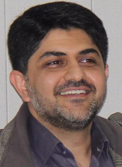
Hossein Rabbani, PhD, SMIEEE
Welcome to my webpage
Here you can find my research/educational activities
About

I received my Msc and PhD in Biomedical Engineering (Bioelectrics) from Amirkabir University of Technology (Tehran Polytechnic) which I was a Visiting Research Scholar in Queen's University for completion of my PhD. I also was a Postdoctoral Research Fellow in Duke University and Postdoctoral Research Scholar in the University of Iowa. Previously, I received a BSc (Hons) in Electrical Engineering (Communications) from Isfahan University of Technology.
You can download my CV.
NEWS

- CV / Resume
- My Google Scholar Page (you can find most papers and cross-references in this link)
- Available Datasets (You can also freely download/upload the datasets from: Isfahan MISP Datasets)
- A summarized Table about ATOMIC Representation
- Dataset, with manual annotations used in our recent automated segmentation of fluorescein leakage in subjects with diabetic macular edema are available here. The slides were uploaded.
- State-of-the-art method for Image Restoration+Codes-->New Codes (The fast version. Thanks Prof. Figueridio for his helps!)
- A summarized Table about Image Modeling
- Statistical Modeling of Retinal Optical Coherence Tomography
- Optical Coherence Tomography Image Analysis (Wiley EEEE Book Chapter)
- Postdoc Position @ MISP (FA)
- Circlelet Transform
- Retinal OCT Classification Challenge (ROCC)
- Slides of Keynote Talk at MVIP2017
- OCT Classification by CNN+Dataset
- Useful Links
Missing Surface Estimation Based on Modified Tikhonov Regularization: Application for Destructed Dental Tissue
DOI: 10.1109/TIP.2018.2800289
IEEE Transactions on Image Processing, 27(5): 2433-2446, 2018.
Research
Field of Interests
Currently I am focusing on Statistical & Mathematical Modeling of Medical Signals and Systems. In this base, my field of interests consists of introducing efficient algorithms for biomedical signal analysis and processing including multidimensional data, time-frequency analysis tools including x-lets, denoising and signal/image recovery, and statistical signal processing.
Learn More12/01/2015MISP research center
Medical Image and Signal Processing (MISP) Research Center is located at the heart of Isfahan University of Medical Sciences. The center was established in 2005 with close collaboration of faculty members from Isfahan University of Medical Sciences and Isfahan university of Technology. At MISP we work on different aspects of biomedical engineering, biomedical image and signal processing. We are dedicated to find new technical solutions for medical devices and fill the gap between the medical and engineering communities. Our missions are To create a place for researchers to work on fundamental, applied and interdisciplinary research projects. To communicate with government in order to contribute to society's health To highlight scientific influence of Isfahan University of Medical Sciences in national and international communities. More information are available at http://misprc.ir.
Learn More03/14/2016Available Datasets
You can freely download/upload the datasets from: Isfahan MISP Datasets (biosigdata.com)
Learn More04/27/2018Optical Coherence Tomography Image Analysis
Check It Out Today!
Wiley EEEE Book Chapter doi.org/10.1002/047134608X.W8315
Course Materials
Fluid/Cyst Segmentation in Optical Coherence Tomography
Check It Out Today!
A. Rashno, D. D. Koozekanani, P. M. Drayna, B. Nazari, S. Sadri, H. Rabbani, K. K. Parhi, "Fully Automated Segmentation of Fluid/Cyst Regions in Optical Coherence Tomography Images With Diabetic Macular Edema Using Neutrosophic Sets and Graph Algorithms," IEEE Transactions on Biomedical Engineering, vol. 65, no. 5, pp. 989-1001, May 2018. A. Rashno, B. Nazari, D. D. Koozekanani, P. M. Drayna, S. Sadri, H. Rabbani, K. K. Parhi, "Fully-automated segmentation of fluid regions in exudative age-related macular degeneration subjects: Kernel graph cut in neutrosophic domain," PLoS ONE 12(10): e0186949. M. Esmaeili, A. Mehri, H. Rabbani, F. Hajizadeh, “Three-dimensional segmentation of retinal cysts from spectral-domain optical coherence tomography images by the use of three-dimensional curvelet based k-SVD ,” Journal of Medical Signals & Sensors, vol. 6, no. 3, pp. 166–171, 2016.
Available Datasets
Dataset for Fluorescein Angiography (Video & Late Image) in DME eyes
The datasets (24 768*768*x FA videos and late FA images in DME eyes) and manual and automated markings used in the following paper can be downloaded from HERE.
OCT data & Color Fundus Images of Left & Right Eyes of 50 healthy persons
This dataset contains OCT data (in mat format) and color fundus data (in jpg format) of left & right eyes of 50 healthy persons.
Bone Marrow Microscopic Data (plasma cell lineage images)
This folder contains bone marrow microscopic images. These images are categorized into two groups: Normal Plasma Cells and Myeloma Cells.
Fundus Fluorescein Angiogram Photographs of Diabetic Patients
We have collected retinal image of 70 patients of different diabetic retinopathy stages including 30 normal data and 40 abnormal data in different stages.
Dataset of Leishmania Parasite in Microscopic Images
45 24-bit 3264*2448 microscopic images taken from bone marrow samples including leshman bodies.
CT & MR Volumes Used for Watermarking of DICOM Images
This dataset contains 260 CT and 202 MR images in DICOM format.
Fundus Fluorescein Angiogram Photographs & Colour Fundus Images of Diabetic Patients
Publicly available database of both fundus fluorescein fngiogram photographs and corresponding color fundus images of 30 healthy persons and 30 patients with diabetic retinopathy.
Database of corneal OCT taken from Heidelberg OCT imaging system (3D .mat data of 15 subjects)
A set of 2D .mat corneal OCT images of 15 subjects. For example subject#1 includes 41 240×748 B-scans taken from Heidelberg OCT imaging device.
Colour Fundus Images of Healthy Persons & Patients with Diabetic Retinopathy
This folder includes 25 colour fundus images of healthy persons and 35 colour fundus images of patients with diabetic retinopathy used for automatic curvelet-based detection of Foveal Avascular Zone (FAZ).
Database of 22 retinal images for the purpose of vessel-based registration of Fundus and OCT projection images of retina
A set of eye images consisting of 22 pairs of images (17 macular and 5 prepapillary), from random patients, each pair acquired from eyes with a variety of retinal diseases. Each image pair includes a colour fundus image and one OCT image acquired with Topcon 3D OCT-1000 instrument. OCT images contain images of 650 different slices with a size of 650 × 512 × 128 voxels and a voxel resolution of 3.125 µm × 3.125 µm × 7 µm.
Red blood cells
A self-provided dataset contains 148 microscopic images of blood smears.
Kidney microscopic images (Glomeruli)
A dataset for Glomeruli detection was collected with the contribution of MISP Research Center and Department of Pathology at IUMS
Dataset for OCT Classification (50 Normal, 48 AMD & 50 DME)
This dataset is acquired at Noor Eye Hospital in Tehran and is consisting of 50 normal, 48 dry AMD, and 50 DME OCTs.
ONH-based OCT of 7 healthy and 7 glaucoma data captured by Heidelberg Spectralis
7 healthy and 7 glaucoma data captured by Heidelberg Spectralis used to demonstrate the efficacy of a new imaging biomarker namely Volumetric Cup-to-Disc Ratio (VCDR) for diagnosis of ocular diseases such as Glaucoma.


